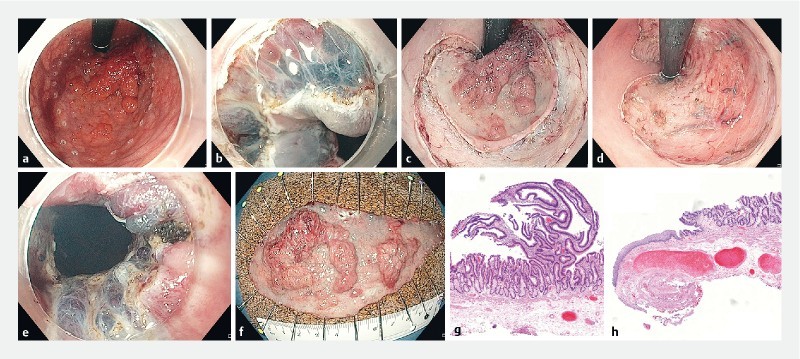
Fig. 1.

ESD of a granular-type LST extending to the dentate line (Paris type 0-IIa + Is). a Retroflexed view after marking the resection borders using hook-knife. b Mucosal incision of the squamous epithelium in the anal canal (forward view). c Retroflexed view after completion of the mucosal incision. d Retroflexed view after completion of ESD. e Distal resection margin including hemorrhoids after completion of ESD (forward view). f Resection specimen (diameter 115 × 50 mm; area 45.1 cm 2 ). g Histopathologic assessment showing adenoma with focal HGIEN resected R0 (H&E, scanning magnification). h Distal resection margin showing R0 resection within the squamous epithelium and thick vessels resected from the rectal venous plexus (H&E, scanning magnification).
