. 2018 Oct 31;27(5):365–376. doi: 10.5607/en.2018.27.5.365

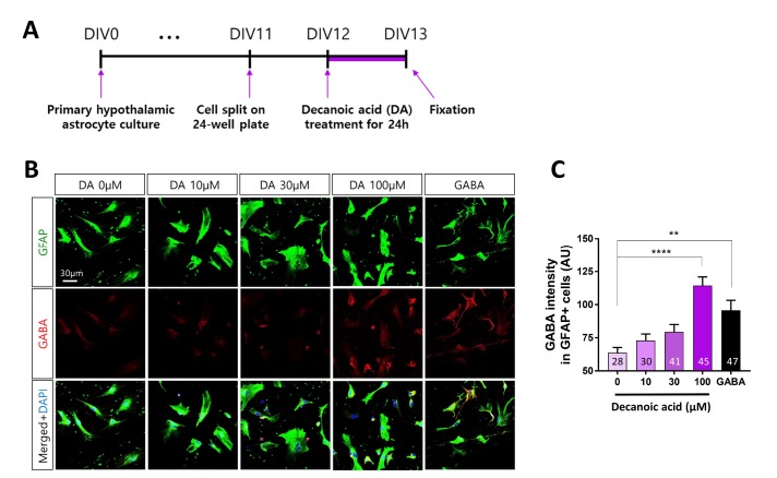
Fig. 5. DA increases GABA production in primary hypothalamic astrocytes. (A) Timeline of treatment of primary hypothalamic astrocytes with different concentrations of DA for 24 h followed by immunocytochemistry. (B) Representative fluorescence immunocytochemical images of cells treated with different concentrations (0, 10, 30, and 100 µM) of DA. Cells were treated with 100 µM GABA as a positive control. (C) Fluorescence intensity of GABA labeling in GFAP-positive primary hypothalamic astrocytes. All data are shown as mean±SEM (0 µM, n=28 cells; 10 µM, n=30 cells; 30 µM, n=41 cells; 100 µM, n=45 cells; and GABA, n=47 cells) and were analyzed using Tukey's multiple comparison test. *p<0.05, **p<0.01, ***p<0.001, ****p<0.0001.

Fig. 5