. 2018 Oct 8;27(5):419–436. doi: 10.5607/en.2018.27.5.419

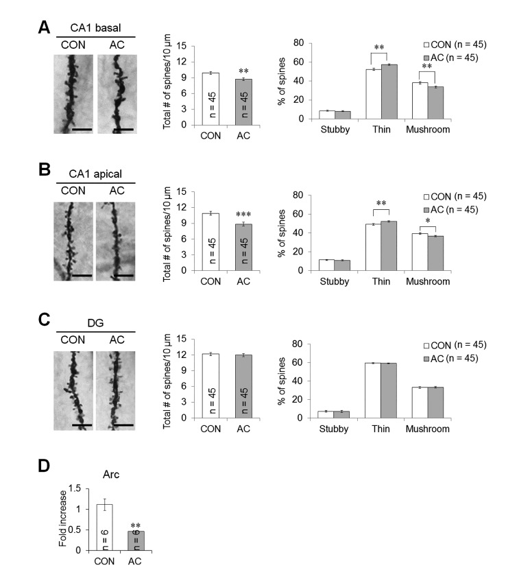
Fig. 5. AC affected dendritic spine density and morphology in the hippocampal CA1 and DG areas. Representative images from each cohort showing dendritic spines along Golgi-impregnated neurons. (A) CA1 basal dendrite. (B) CA1 apical dendrite. (C) DG dendrite. Bar graphs (middle) showed the mean total number of spines, indicating that AC reduced spine density in the CA1 dendritic area. Bar graphs (right) showed the mean proportion of spine morphological subtypes, indicating that AC reduced the proportion of mature spines in the CA1 dendritic area. (D) Changes in the mRNA expression of Arc. The level of Arc mRNA significantly reduced following chronic AC treatment. Data are expressed as means±SEs. *p<0.05, **p<0.01, and ***p<0.001 vs. vehicle-treated controls. Scale bars in the dendritic images represent 5 µm.

Fig. 5