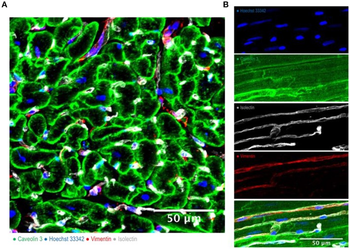
Figure 1.
Cardiac multicellularity in vitro: Immunohistochemicalstaining and confocal microscopywere used to identify cardiac cells in a transverse section (A) and in a longitudinal section (B) of freshly prepared dog myocardial slices. Cardiomyocytes were labeled with caveolin 3, fibroblasts were labeled with vimentin and endothelial cells were labeled with isolectin. Nuclei were labeled with Hoechst 33342. Scale bar = 50 μm.

