Figure 1.
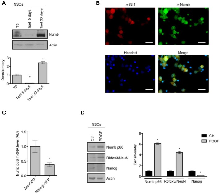
Numb expression in NSCs. (A) Representative Western Blot (WB) (up) and densitometric analysis (bottom) of endogenous Numb isoforms (p66 and p72) in NSCs grown in selective medium for 5 or 30 days, compared to bulk cells (T0). Actin was used as loading control. P-values *p < 0.05. (B) Representative images of immunofluorescence staining of NSCs for Numb (green) and Gli1 (red); nuclei are counterstained with Hoechst (blue). Scale bar: 10 μm for all panels. (C) qRT-PCR analysis showed mRNA expression of Numb p66, evaluated in NSCs infected with lentivirus carrying Nanog-GFP or Zeo-GFP as control and sorted for GFP. Data represent means ± SD from three independent experiments. P-values *p < 0.05. (D) Representative Western Blot (WB) (left) and densitometric analysis (right) of NSCs before and after in vitro differentiation for 48 h (PDGF); p66, Rbfox3/NeuN, Nanog were evaluated. Actin was used as loading control. P-values *p < 0.05. (A–D) Data are means ± SD from three independent experiments. Full-length images are presented in Supplementary Figures.

