Figure 3.
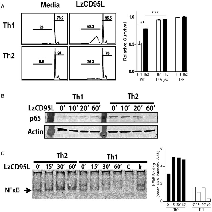
Th2 cells preferentially signal through non-apoptotic Fas pathways following FasL treatment. In vitro skewed Th1 and Th2 cells from WT, LPRcg/wt, and LPR mice were treated with LzCD95L and assayed for induction of apoptosis 4 h later by propidium iodide staining. (A) Representative PI staining from WT Th1 and Th2 cells showing apoptotic cells in sub-G1, and relative survival rate of T cells from different mice following LzCD95L treatment. (B) WT Th1 and Th2 cells were treated with LzCD95L and assayed for cytoplasmic levels of p65 by western blot. (C) WT Th1 and Th2 cells were treated with LzCD95L and assayed for nuclear NFκB binding activity by EMSA. Densitometry measurements (right) are displayed as mean pixel intensities in arbitrary units. Data are representative data from three or more independent experiments. In panel (A) apoptosis assays included 5 replicated for each independent experiment (**p ≤ 0.01, ***p ≤ 0.001).

