
Figure 1.
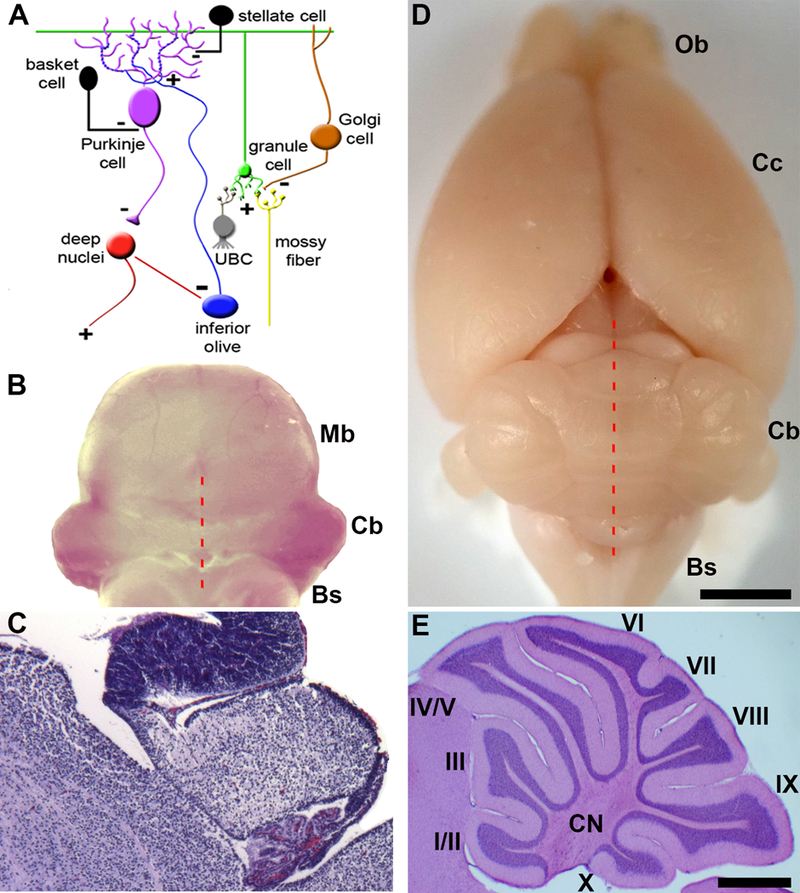
Connectivity and gross morphology of the cerebellum. A) Cellular composition and circuitry of the cerebellum. (+) indicates excitatory synapses and (−) indicates inhibitory synapses. B) Poster view of an embryonic day 16 mouse hindbrain. C) Sagittal tissues section of an embryonic day 16 mouse cerebellum stained with H&E. Provided are general anatomical landmarks for orientation with respect to the cerebellum, with the choroid plexus located posteriorly, the midbrain anteriorly, and the brainstem ventrally. D) Dorsal view of an adult mouse brain. The red broken lines in panels B and C indicate the midline of the cerebellum. E) Sagittal section of the mouse cerebellum showing the structure of the lobules. The lobules are labeled with Roman numerals. Abbreviations: UBC = unipolar brush cell, Bs = brainstem, Cb = cerebellum, Cc = cerebral cortex, CN = cerebellar nuclei, Cp = choroid plexus, fl/pfl = flocculus/paraflocculus, gl = granular layer, Mb = midbrain, ml = molecular layer, Ob = olfactory bulb, V= vermis, H = hemisphere. Scale bar in D = 2mm (applies to B where it = 1mm) and the scale bar in E = 500μm (applies to C where it = 200μm). Panel A was reused with permission from Reeber et al. (2013; Frontiers in Syst Neurosci, 7:83).