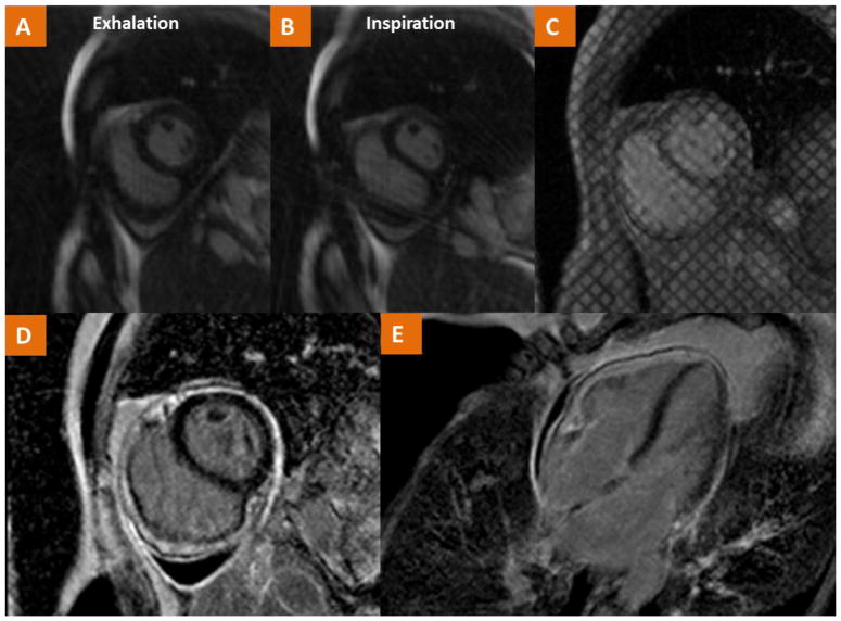
Fig. 8.
CMR findings in a case of a 54 year old female with history of Hodgkin’s Lymphoma treated with radiation at ages 20, 24, 27 and now with recurrent pericardial effusion and dyspnea. (A–B) Real-time cine imaging demonstrates flattening of the septum during inspiration consistent with ventricular interdependence as well as a small pericardial effusion. (C) Tagged cine gradient imaging used to detect fibrotic adhesion of pericardial layers. (D–E) LGE imaging demonstrates pericardial LGE consistent with pericardial inflammation. The consolidation of these findings is consistent with the diagnosis of pericardial constriction.

