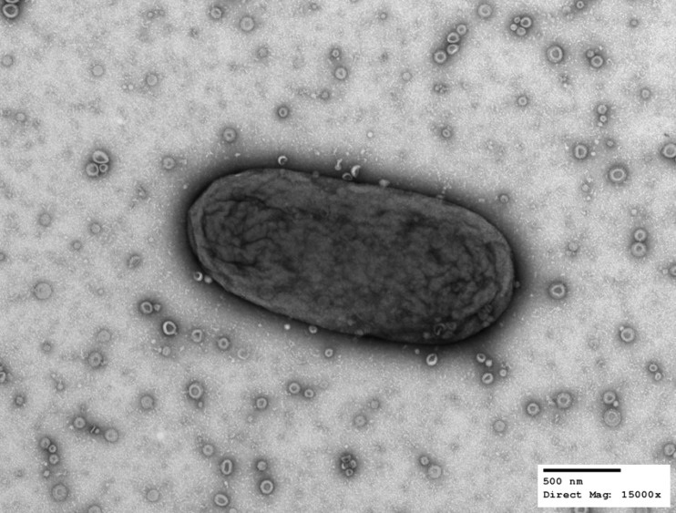
FIG 1.
B. thetaiotaomicron produces outer membrane vesicles (OMVs). Transmission electron microscopy of a single B. thetaiotaomicron cell together with OMVs in the extracellular milieu. B. thetaiotaomicron cells were swabbed from a solid-medium plate, suspended in PBS, and processed for TEM. Images were acquired at a direct magnification of ×15,000. The scale bar represents 500 nm. (Image courtesy of Wandy Beatty, Molecular Microbiology Imaging Facility, WUSTL.)

