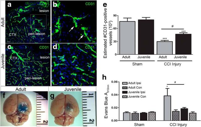
Figure 5.
Vascular density and BBB stability is enhanced in juvenile mice following CCI injury. a, Representative low-magnification confocal image of DAPI (blue) and CD31+ vessels (green) in the perilesion cortex at 4 d post-CCI injury in adult mice. b, Representative high-magnification confocal image from image a inset. c, Low-magnification confocal image of CD31+ vessels in the perilesion cortex at 4 d post-CCI injury in adult mice. d, High-magnification confocal image from image c, inset. e, Quantification of the number of CD31+ vessels by nonbiased stereology shows a significant decrease in the number of vessels in adult and juvenile ipsilateral cortex compared with their shams (***p < 0.001 and ***p = 0.0007, respectively). Juvenile injured mice displayed an increase in CD31+ vessels compared with adult injured (#p = 0.0292). f, Representative image of Evans blue in the adult cortex at 4 d post-CCI injury. g, Representative image of Evans blue in the juvenile cortex at 4 d post-CCI injury. h, Quantification of Evans blue A610nm showing a significant increase in the adult ipsilateral injured cortex at 4 d compared with ipsilateral sham injury or contralateral (Con) hemispheres (*p = 0.0433). Juvenile mice showed a significant reduction in Evans blue absorbance in the ipsilateral cortex compared with ipsilateral adult cortex (#p = 0.0281). Scale bar, 20 μm in b and d and 100 μm in a and c.

