Figure 9.

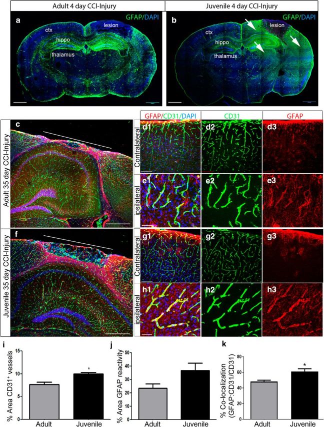
Prominent GFAP immunoreactivity accompanies neuroprotection in juvenile mice after CCI injury. a, Representative mosaic confocal image of GFAP colabeling at 4 d post-CCI injury. b, Representative mosaic confocal image of GFAP colabeling at 4 d post-CCI injury in juvenile mice. Increased immunoreactivity for GFAP is seen in the perilesion cortex, hippocampus, and thalamus (white arrows) compared with adult. c, Confocal mosaic image of the ipsilateral cortex at 35 d post-CCI injury in adult mice. Double immunolabeling for GFAP (red) and CD31 (green) shows the glial scar in the perilesion cortex. d1–d3, High-magnification confocal image of GFAP and CD31 in contralateral and ipsilateral cortex (e1–e3) after CCI injury. f, Confocal image of the juvenile ipsilateral cortex at 35 d post-CCI injury. High-magnification confocal image of GFAP and CD31 in contralateral (g1–g3) and ipsilateral (h1–h3). i, Quantification of the percentage area of CD31+ vessels in the perilesion at 35 d shows an increase in vessel density in the juvenile perilesion compared with adult mice (*p = 0.0143). No difference was seen in the percentage area of GFAP reactivity (j), but a significant increase in the percentage colocalization was seen (GFAP:CD31/CD31) (*p = 0.0332) (k). ctx, Cortex; hippo, hippocampus. Scale bar, 1 mm in a–c, and f; 500 μm in d1 and g1; and 100 μm in e1 and h1.
