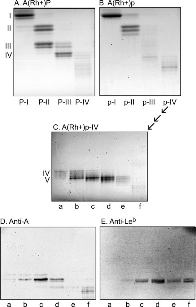
Figure 3.

Glycosphingolipid subfractions isolated from human blood group A(Rh+)P and A(Rh+)p stomachs. A–E, thin-layer chromatograms after detection with anisaldehyde (A–C) and autoradiograms obtained by binding of monoclonal antibodies directed against the blood group A determinant (D) and the Leb determinant (E). The glycosphingolipids (4 μg/lane) were separated on glass or aluminum-backed silica gel plates, using chloroform/methanol/water (60:35:8 by volume) as the solvent system, and the binding assays were performed as described under “Experimental procedures.” Autoradiography was for 12 h. The Roman numerals to the left of A and C denote the number of carbohydrates in the bands.
