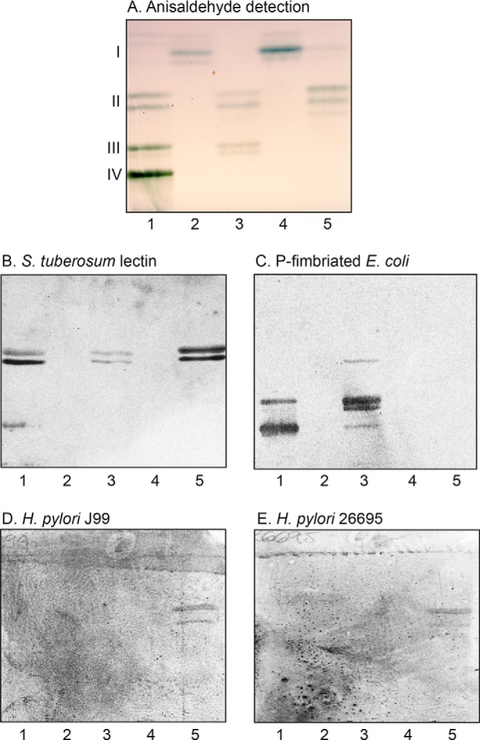
Figure 4.

Binding of H. pylori, P-fimbriated E. coli, and S. tuberosum lectin to mono-, di-, and triglycosylceramides of human stomach. A–E, thin-layer chromatograms of the mono- to triglycosylceramide fractions isolated from human A(Rh+)P and A(Rh+)p stomachs (A) and autoradiograms obtained by binding of S. tuberosum lectin (B), P-fimbriated E. coli strain 291-15 (C), H. pylori strain J99 (D), and H. pylori strain 26695 (E). The glycosphingolipids were separated on aluminum-backed silica gel plates, using chloroform/methanol/water 65:25:4 (by volume) as the solvent system, and the binding assays were performed as described under “Experimental procedures.” Autoradiography was for 12 h. Lane 1, reference nonacid glycosphingolipids of human blood group O erythrocytes, 40 μg; lane 2, fraction P-I, 4 μg; lane 3, fraction P-II, 4 μg; lane 4, fraction p-I, 4 μg; lane 5, fraction p-II, 4 μg. The Roman numerals to the left of the chromatogram in A denote the number of carbohydrate units in the bands.
