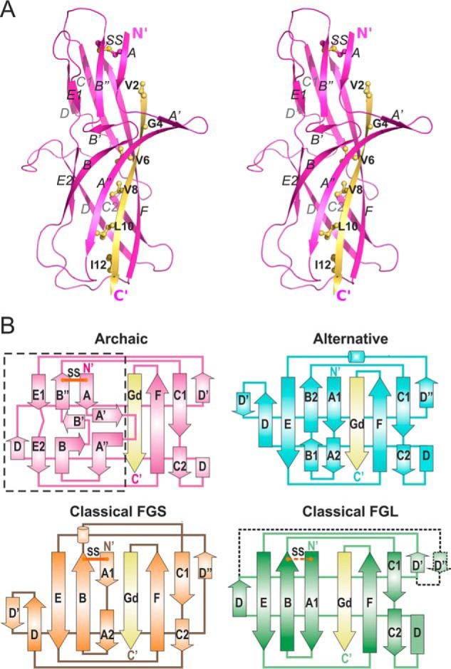
Figure 2.

The first β-sheet of CsuA/B has a unique, non-Ig-like architecture. A, crystal structure of CsuA/Bsc (cartoon representation, stereo view). The Gd donor strand is colored yellow and donor residues are shown as balls-and-sticks and labeled. B, topology diagrams of pilins from archaic, classical FGS chaperone assembled, classical FGL chaperone assembled, and alternative CUP systems. Donor strands are shown in yellow. Positions of conserved disulfide bonds (SS) are shown with lines in red. In the classical FGL subfamily, the disulfide bond and additional D″ strand are present in some subunits and absent in others, and hence are shown using dashed lines. The unique for archaic systems β-sheet ABE is framed in a rectangle. Superpositions of pilins from different CUPs are shown in Fig. S4.
