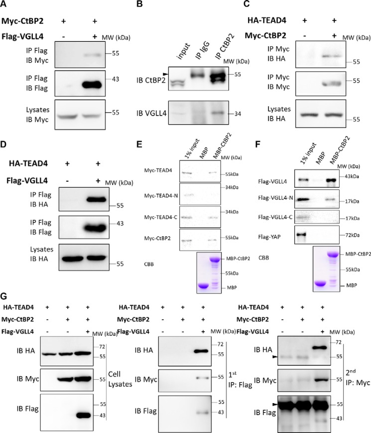
Figure 4.
CtBP2 formed a complex with TEAD4-VGLL4. A, co-IP assay of the interaction between CtBP2 and VGLL4. The indicated plasmids were transfected into 293T cells and analyzed by co-IP. IB, immunoblot; MW, molecular weight. B, the endogenous interaction between CtBP2 and VGLL4. 293T cells lysates were incubated with CtBP2 or IgG antibodies, followed by immunoprecipitation and Western blot analysis with the indicated antibodies. The arrowhead indicates the IgG heavy chain. C and D, co-IP assay of the interaction between TEAD4 and CtBP2 (C) and TEAD4 and VGLL4 (D). The indicated plasmids were transfected into 293T cells and analyzed by co-IP. E, an MBP pulldown assay showed that the TEAD4 and TEAD4 C termini were pulled down by MBP-CtBP2. MBP or MBP-CtBP2 bound to MBP beads were incubated with IVTT 35S-labeled TEAD4/TEAD4-N/TEAD4-C for 3 h. Immobilized complexes were then washed and subjected to SDS-PAGE. The input was 10% of the total amount of IVTT 35S-TEAD4/TEAD4-N/TEAD4-C. CBB, Coomassie Brilliant Blue staining. F, an MBP pulldown assay showed that VGLL4 and VGLL4 N termini were pulled down by MBP-CtBP2. MBP or MBP-CtBP2 bound to MBP beads were incubated with IVTT 35S-labeled VGLL4/VGLL4-N/VGLL4-C for 3 h. Immobilized complexes were then washed and subjected to SDS-PAGE. The input was 10% of the total amount of IVTT 35S-VGLL4/VGLL4-N/VGLL4-C. G, 293T cells expressing the indicated proteins were collected for two-step immunoprecipitation and analyzed by Western blotting. Cell lysates were first co-immunoprecipitated with M2-FLAG beads and then eluted for a second immunoprecipitation with Myc antibody. The arrowheads indicate the IgG heavy chains.

