Figure 2.
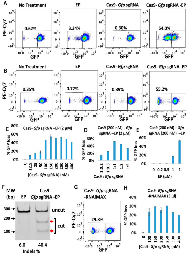
Efficient gene deletion achieved in GFP-J774A.1 cells following treatment with CriPs targeting Gfp. At 48 h or at 5 days post-treatment, flow cytometry and T7E1 assays were performed to measure the loss of GFP. A, 48 h post-treatment, GFP loss measurements by flow cytometry. B, 5 days post-treatment, GFP loss measurements by flow cytometry. Concentrations were as follows: Cas9, 150 nm; Gfp sgRNA, 150 nm; EP, 2 μm. C, different concentrations of Cas9–Gfp sgRNA (1:1) with 2 μm EP. D, different ratios of Cas9 (200 nm) to Gfp sgRNA with 2 μm EP. E, different concentrations of EP with 200 nm Cas9–Gfp sgRNA. F, percentage indel measurements in Gfp genomic DNA isolated from CriP-treated cells versus EP-treated cells by a T7E1 assay (uncut, 292 bp; cut, 179 bp + 113 bp; Cas9, 200 nm; Gfp sgRNA, 200 nm; EP, 2 μm). G, flow cytometry data of GFP-expressing J774A.1 cells treated with RNAiMAX-mediated delivery of Cas9–Gfp sgRNA at 5 days post-treatment (Cas9, 150 nm; Gfp sgRNA, 150 nm). H, different concentrations of Cas9–Gfp sgRNA (1:1) with RNAiMAX (3 μl). Error bars, S.E.

