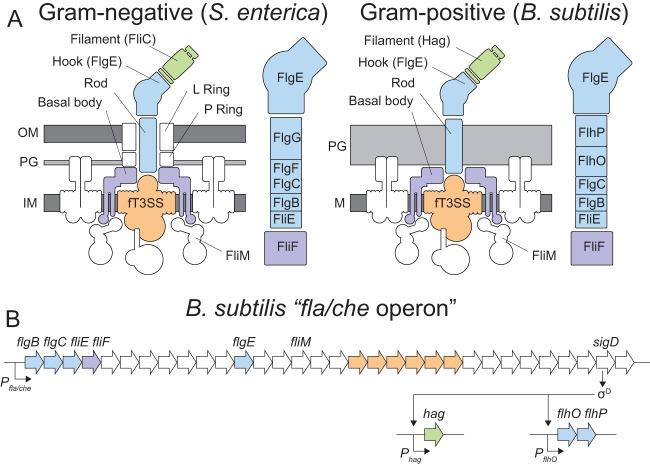
FIG 1.
Diagrams of B. subtilis flagellar structure and genetic architecture. (A) Cartoon model of the Gram-negative and Gram-positive flagellar architectures. To the right of each flagellum is a closeup depiction of the assembly order of the rod components (14). The position of FlgC in the Gram-negative rod has traditionally been considered cell proximal to FlgF, but a recent publication suggests that the orders may be reversed (25). The order of the rod components in B. subtilis is indicated based on the information in the present manuscript. The membrane is colored dark gray. Peptidoglycans are colored light gray. The flagellar basal body is colored purple, the flagellar type III secretion system (fT3SS) is colored orange, the rod-hook structure is colored blue, and the filament is colored green. (B) B. subtilis fla/che operon structure and flagellar genetic hierarchy. Genes mentioned in the manuscript are indicated in italics. Bent arrows indicate promoters, and open arrows indicate genes. Genes are color coded to match the relative locations of their gene products in panel A.

