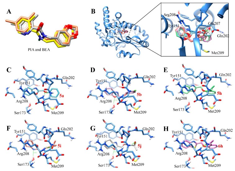
Figure 4.
The binding models of the test compounds into the lipid binding pocket of Sec14p from S. cerevisiae. (A) Superposition of PIA (yellow) and BEA (orange) in docking resultant models with PIA in the X-ray crystallographic structure (PDB 6F0E; gray). (B) Overlay of the six test benzoxazole and benzothiazole derivatives. (C–H) The binding modes of the compounds (in stick model with carbon) into the active site of Sec14p: (C) 5a (aquamarine); (D) 5b (orchid); (E) 5h (light green); (F) 5i (coral); (G) 5j (light gray); and (H) 6h (hot pink).

