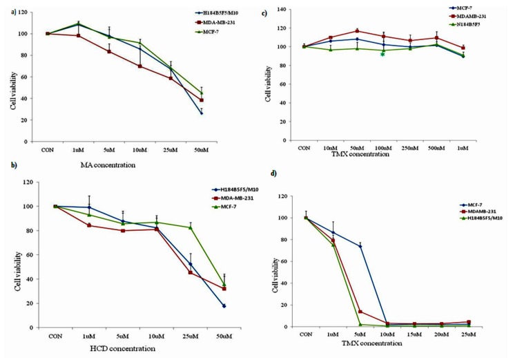
Figure 1.
Effect of MA, HCD, and TMX on cell viability of breast cancer cell lines and human mammary epithelial cells by MTT assay. The viability of all the three cell lines was measured after a 24-h treatment with the indicated concentrations of (a) MA (1, 5, 10, 25, and 50 µM); (b) HCD (1, 5, 10, 25, and 50 µM); (c) low dose of Tamoxifen (10, 50, 100, 250, 500, and 1000 nM); and (d) high dose of Tamoxifen (1, 5, 10, 15, 25, and 25 μM). Data shown are the mean values ± SE from three independent experiments. Statistical analyses were determined using one-way ANOVA followed by post hoc Tukey multiple comparison test with ** p < 0.01, *** p < 0.001, significantly different from control.

