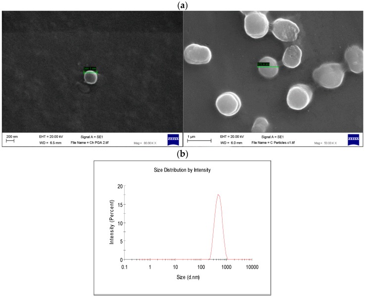
Figure 7.
(a) Scanning electron microscopy (SEM) images of γ-PGA nanoparticles (NPs); (b) Dynamic light-scattering (DLS) analysis of γ-PGA NP formed by ionic gelation method; γ-PGA NPs formed by the formulation of (0.05:0.15 CH:γ-PGA w/v). Magnification power is 80,000× (left image) and 50,000× (right image).

