Figure 2.
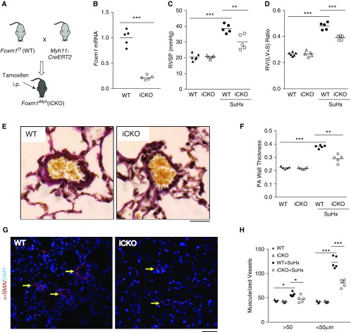
Foxm1 disruption in smooth muscle cells (SMCs) inhibits Sugen 5416/hypoxia (SuHx)-induced pulmonary hypertension in mice. (A) A diagram showing the strategy for generating tamoxifen-inducible SMC-specific knockdown of Foxm1 in mice (iCKO). (B) Tamoxifen treatment induced loss of FoxM1 expression in SMCs (α-SMA+) isolated from iCKO mice compared with wild-type (WT) mice. (C) Hemodynamic measurement showing that iCKO mice had decreased right ventricular systolic pressure compared with Foxm1f/f (WT) mice after SuHx treatment. (D) Attenuation of RV hypertrophy in iCKO mice compared with WT mice after SuHx treatment. (E) Representative micrographs of Russel-Movat pentachrome staining showing increased medial thickness in SuHx WT mice compared with SuHx iCKO mice. (F) Quantification of pulmonary artery wall thickness. Pulmonary arteries with a diameter of 20–200 μm were quantified. (G and H) Muscularization of distal pulmonary vessels was markedly inhibited in SuHx iCKO mice compared with SuHx WT mice. Lung sections were immunostained with anti–α-SMA (red). Arrows indicate α-SMA+ distal pulmonary vessels. α-SMA+ vessels were quantified in 100 fields per sample. *P < 0.05, **P < 0.01, and ***P < 0.001. (B) Student’s t test. (C, D, F, and H) One-way ANOVA. Horizontal bars in dot plot panels represent the mean. Scale bars: (E) 100 μm; (G) 50 μm. Foxm1 = forkhead box M1; PA = pulmonary artery; RV/(LV + S) = right ventricle versus left ventricle plus septum; RVSP = right ventricular systolic pressure; SMA = smooth muscle actin.

