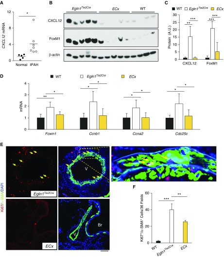
Figure 5.
Role of endothelial Cxcl12 in activating FoxM1 (forkhead box M1)-dependent smooth muscle cell proliferation in vivo. (A) qRT-PCR analysis demonstrating increased CXCL12 expression in lung tissues of patients with IPAH compared with healthy donors. (B and C) Western blotting and densitometric analysis demonstrating inhibited FoxM1 expression in the lungs of Egln1/Cxcl12Tie2Cre double-knockout mice (ECx) compared with Egln1Tie2Cre mice. At 3.5 months of age, mouse lungs were collected for Western blotting analysis. These data demonstrated that endothelial cell–derived Cxcl12 secondary to Egln1 deficiency induced FoxM1 expression in vivo. (D) qRT-PCR analysis showing normalization of FoxM1 and its downstream genes essential for cell cycle progression, Ccnb1, Ccna2, and Cdc25c in ECx lungs compared with Egln1Tie2Cre lungs. n = 4 wild-type, 5 Egln1Tie2Cre, and 5 ECx. (E and F) In vivo cell proliferation assessment by anti-Ki67 immunostaining showing decreased smooth muscle cell proliferation in ECx lungs compared with Egln1Tie2Cre lungs. Lung cryosections were immunostained with anti-Ki67 (red) and anti–α-smooth muscle actin (green). Nuclei were counterstained with DAPI. Arrows point to proliferating smooth muscle cells. n = 4/each group. *P < 0.05, **P < 0.01, and ***P < 0.001. (A) Welch t test. (C, D, and F) One-way ANOVA. Scale bar, 50 μm. A.U. = arbitrary units; Br = bronchiole; IPAH = idiopathic pulmonary arterial hypertension; SMA = smooth muscle actin; V = vessel; WT = wild type.

