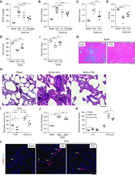
Figure 7.
Thiostrepton (Thio) inhibition of FoxM1 (forkhead box M1) inhibits pulmonary hypertension in Sugen 5416/hypoxia (SuHx) rats. (A) Reduction of right ventricular systolic pressure by Thio treatment in SuHx rats compared with vehicle (Veh)-treated SuHx rats. Three weeks post-SuHx challenge, the rats were returned to normoxia and treated with Thio (10 mg/kg, or 20 mg/kg, i.p. daily) or DMSO (Veh) for 3 weeks. (B) Right ventricle (RV) hypertrophy was markedly decreased by higher dose of Thio (20 mg/kg) treatment in SuHx rats. (C–F) Echocardiography revealing reduced RV wall thickness, improved RV contractility, and enhanced cardiac output and pulmonary artery (PA) diastolic function as evidenced by normalization of pulmonary arterial acceleration time/ejection time ratio in Thio-treated SuHx rats compared with respective control animals. Thio, 20 mg/kg treatment. (G) Trichrome staining demonstrating marked inhibition of RV fibrosis (blue) by Thio treatment in SuHx rats. (H–J) Russel-Movat pentachrome staining demonstrating reduced PA occlusion lesions and wall thickness. Arrows indicate occlusive vascular lesions. PAs in a diameter of 20–200 μm were quantified. (K and L) Reduction of distal pulmonary vessel muscularization by Thio treatment. Sections of rat lung tissues immunostained with anti–α-smooth muscle actin antibody (red). Nuclei counterstained with DAPI (blue). Arrows indicate muscularized distal vessels. *P < 0.05, **P < 0.01, and ***P < 0.001. (A–F, J, and K) One-way ANOVA. (I) Student’s t test. Scale bars: (G) 100 μm; (H and L) 50 μm. CO = cardiac output; n.s. = not significant; PA AT/ET = pulmonary arterial acceleration time/ejection time; RVFAC = right ventricular fractional area change; RVWTD = right ventricle wall thickness in diastole; RV/(LV + S) = right ventricle versus left ventricle plus septum; RVSP = right ventricular systolic pressure; SMA = smooth muscle actin.

