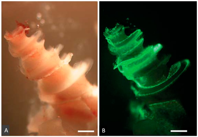
Figure 2.
Photomicrographs of whole-mounted guinea pig cochlea showing the distribution of GFP-fusion protein after injection of GFP-fused Sendai virus vector (GFP-SeV/ΔF) into the scala media. Light (A) and fluorescent (B) images of the cochlea of a SeV-inoculated ear. Scale bars: 1000 μm (This figure was cited by reference [10] and permitted by S. Karger AG, Medical and Scientific Publishers.).

