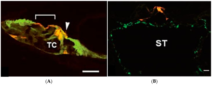
Figure 3.
Photomicrographs of histological sections of guinea pig scala media after injection of GFP-SeV/ΔF (A) and after injection via a scala tympani approach (B). (A) Hair cells and supporting cells from the organ of Corti in an inoculated ear. An inner hair cell is indicated by an arrowhead, and an outer hair cell region is delimited by a bracket. Red, F-actin-stained with rhodamine phalloidin; green, GFP-SeV/ΔF-transfected cells. Scale bars: 10 μm. (B) Sensory epithelial cells and fibrocytes of the scala tympani. Numerous fibrocytes in the scala tympani are fluorescently labeled. BM, basilar membrane; SL, spiral limb; ST, scala tympani; TC, tunnel of Corti. Scale bars: 100 μm. (This figure was cited by reference [10] and permitted by S. Karger AG, Medical and Scientific Publishers.)

