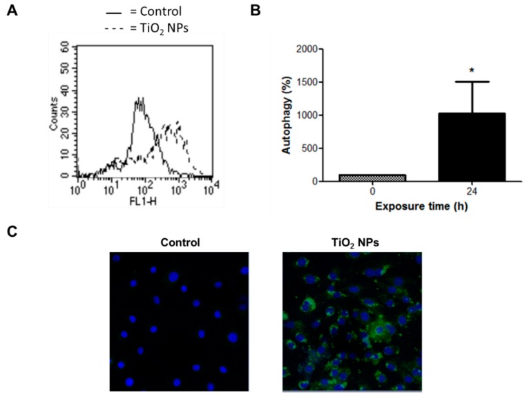
Figure 8.
TiO2 NPs induced autophagy. H9c2 cells were treated with 20 μg/cm2 TiO2 NPs for 24 h and autophagy was evaluated through a detection kit by flow cytometry (A,B) and confocal microscopy (C). In (B), results are presented as mean ± standard deviation (SD) of three independent experiments (n = 3). * Significant difference between untreated cells (0) and TiO2 NPs-treated cells (p < 0.05). In (C), nuclear stain with DAPI and green detection reagent (autophagy) are showed.

