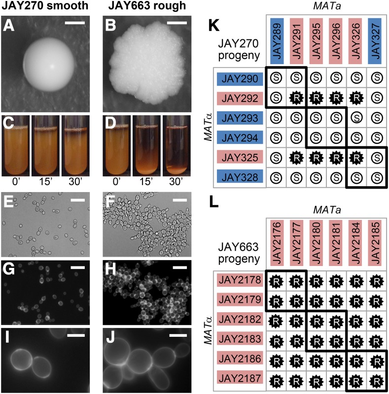
Figure 1.
Smooth and rough colony morphologies, liquid sedimentation, mother-daughter cell attachment, and phenotypes of diploids derived from mating specific haploids. A-J show images of the JAY270 smooth parent diploid strain (left panels) and its spontaneous rough derivative JAY663 (right panels). A and B, colony morphologies on YPD agar after 3 days growth at 30C. C and D, cell sedimentation kinetics. 5 ml liquid YPD cultures were grown overnight at 30C in a rotating drum. Test tubes were vortexed vigorously for 10 sec to fully resuspend the cells, then were left to rest and photographed at 15 min intervals (the 0’ pictures were taken immediately after vortexing). E and F, bright field, and G-J, fluorescence microscopy of cells stained with calcofluor white to highlight chitin septa and the mother-daughter cell attachment. Scale bars are 1mm (A-B), 20µm (E-F) and 5µm (G-J). K and L, Smooth (S, white circles) and rough (R, black stars) phenotypes of diploids formed by crossing the indicated MATa and MATα haploids isolated from three tetrads of each JAY270 (K) and JAY663 (L). Thick black lines indicate the four diploids derived from matings of intra-tetrad sibling haploids. The colored backgrounds for each haploid correspond to their inferred genotype (Blue, dominant wild type allele; Red, recessive mutant allele). All 12 haploids from panel K had their whole genomes sequenced. Co-segregation analysis with JAY270 HetSNPs (Fig. S1) was used for identification of the causal mutation at the ACE2 locus (Fig. 2).

