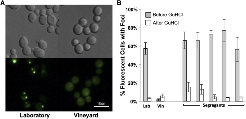
Figure 1.
Rnq1 prion modifies Htt-Q75-GFP protein aggregation. (A) Laboratory and vineyard parent strains induced to express the Rnq1-YFP fusion protein for 12 h were imaged. Representative images of DIC (top) and fluorescent patterns of the Rnq1-YFP protein (bottom) are shown. Focal YFP signal present in the Laboratory strain images indicate [PIN+] cells while diffuse YFP signal in the vineyard strain indicates [pin-] cells. (B) Aggregation of the Htt-Q75-GFP protein was quantitated in Laboratory (Lab), Vineyard (Vin) parent strains and five arbitrarily selected segregants before and after successive passaging on GuHCl plates to eliminate endogenous prions. Aggregation is represented as the percentage of GFP-fluorescent cells with foci.

