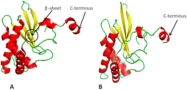
Figure 4.
(A) LuxS homology model and (B) predicted 3D-structural model of the LuxS protein. Red indicates alpha helices, yellow indicates sheets, and green indicates loops. The difference between template and predicted model is found. The template contains one extra beta sheet shown within the black circle (about two residues long) and has long alpha helices. The predicted model at C-terminus alpha helix has short, long loops and one residue found is the loop at C-terminus, while the template does not contain any loop residue at the C-terminus.

