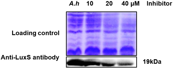
Figure 8.
Validation of LuxS protein expression levels with Western blotting using increasing concentrations of a LuxS inhibitor compound. A. hydrophila was treated and untreated (A.h, wild type used as a control) with increasing inhibitor concentrations of 10, 20, and 40 μM (lower panel). Coomassie R-350 staining of the membrane shows equal loading of the protein sample (upper panel).


