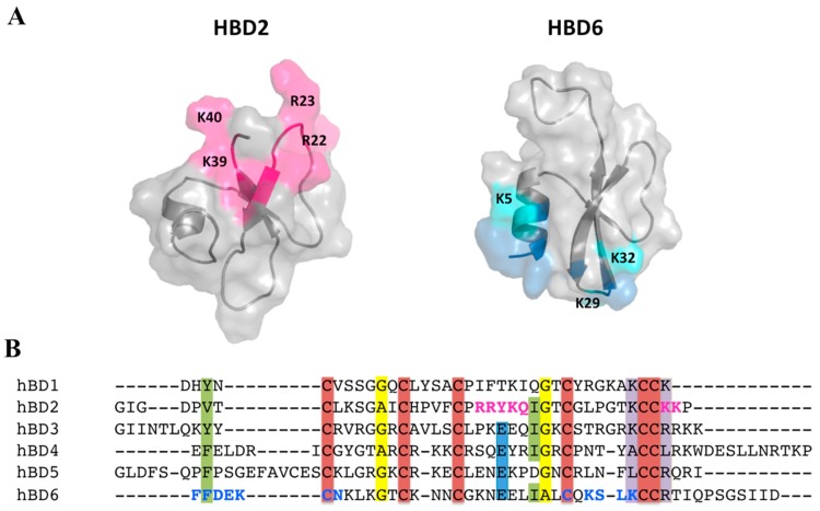
Figure 5.
β-Defensin interaction with glycosaminoglycan (GAG), mapped using nuclear magnetic resonance (NMR) chemical shift perturbation (CSP). (A) Comparison of the heparin-binding motif mapped on the surfaces of human β-defensin (HBD)2 and HBD6. Residues exhibiting pronounced NMR CSP upon GAG binding are highlighted in magenta for HBD2 [53] and blue for HBD6 [41] on the surface and in the primary sequence. The CSPs for HBD2 were reproduced from [53]. (B) Sequence alignments for six HBDs, performed using ClustalW software. Protein names are indicated on the left; amino acids are color-coded based on conservation scores 9, calculated using the ConSurf server. Highly conserved Cys, Gly, Glu, and basic residues are shown in red, yellow, blue, and purple backgrounds, respectively.

