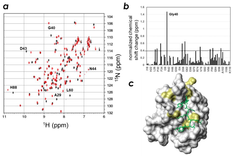
Figure 4.
Identification of the key residues of the mouse ZO-1(PDZ1) domain responsible for CLD-3 peptide recognition. (a) Overlaid spectra of mouse ZO-1(PDZ1) in the presence (red) and absence (black) of the CLD-3 peptide. (b) Normalized chemical shift changes of mouse ZO-1(PDZ1) domain upon mixing with the 2 equivalence of the CLD-3 peptide. (c) Resonances representing residues with larger chemical shift changes than the threshold values are mapped onto a van der Waals surface diagram of the mouse ZO-1(PDZ1) (PDB: 2rrm) and displayed in yellow. The threshold value is indicated by the dashed line in graph (b).

