Figure 6.

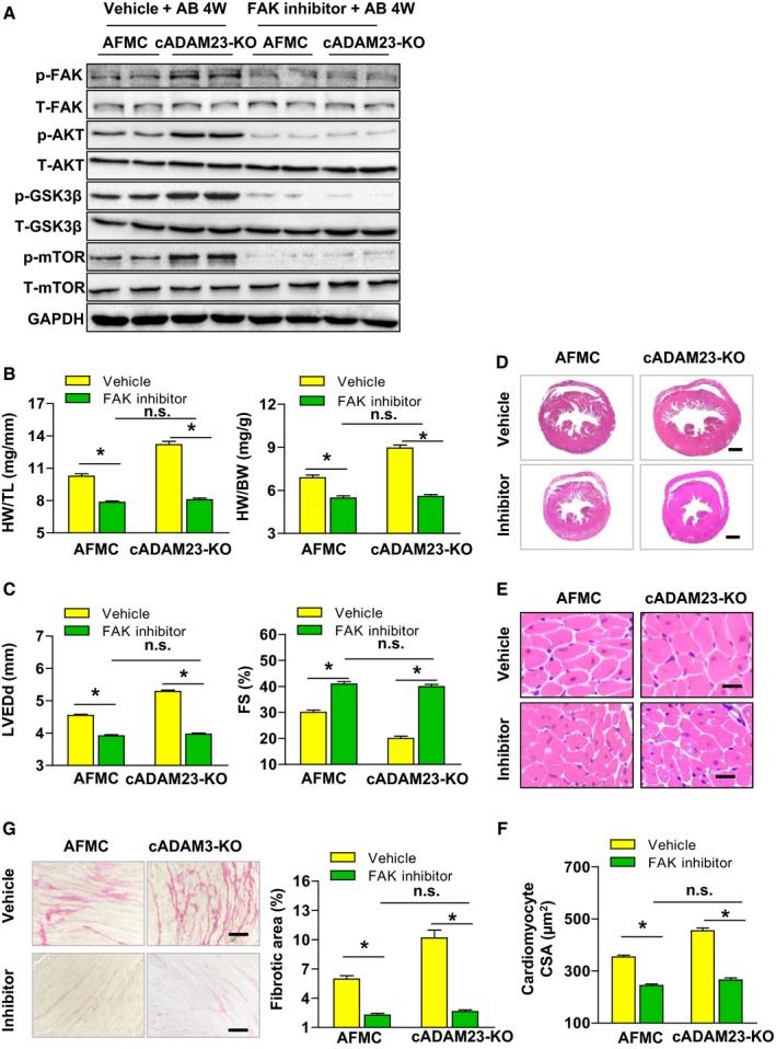
FAK inhibitor blunted aortic banding (AB)‐induced cardiac hypertrophy in cADAM23‐KO mice in vivo. A, AFMC and cADAM23‐KO mice were treated with FAK inhibitor (PF‐562271) or vehicle for control after 4 weeks of AB surgery. Western blot analysis and statistical results of the phosphorylation and total protein levels of FAK, AKT, m‐TOR and GSK3β in heart tissues in the indicated groups (n=4 per group). B, Statistical results for HW/BW, and HW/TL ratios (n=12 mice per group). C, Statistical results for the echocardiographic parameters LVEDd and FS in indicated groups (n=12 mice for AFMC vehicle group, n=11 mice for cADAM23‐KO vehicle group and cADAM23‐KO inhibitor group, n=10 mice for AFMC inhibitor group). D and E, Representative images of the histological analysis of cardiac hypertrophy, as indicated by the whole heart (D) and heart sections (E) stained with H&E in cADAM23‐KO and AFMC mice treated with PF‐562271 or vehicle (for D, scale bars, 1 mm; for E, scale bar, 20 μm). F, Statistical results for the cardiomyocyte CSA in the indicated groups (n>100 cells per group). G, Representative images of cardiac fibrosis and statistical results for the fibrotic area (n>40 fields per group, scale bar, 20 μm). For (B, C, F, and G), CSA indicates cross‐sectional area, FAK, focal adhesion kinase; H&E, hematoxylin and eosin; HW/BW, heart weight to body weight; HW/TL, heart weight to tibia length; LVEDd, left ventricular end‐diastolic diameter; NRCM, neonatal rat cardiomyocytes; n.s., no significance. *P<0.05 vs vehicle.
