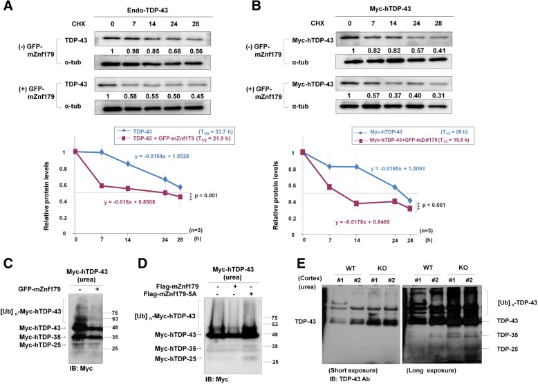
Fig. 6.
Znf179 enhances the degradation rate of TDP-43 protein and alters the solubility of TDP-43. a and b Endogenous TDP-43 (a) or Myc-hTDP-43 (b) in N2a cells with or without stably expressed GFP-mZnf179 was treated with cycloheximide (20 mg/ml) for different time periods. The protein levels of the Myc-hTDP-43 were analyzed by western blotting with anti-TDP-43 or anti-Myc antibody. The graph below the blots showed a quantification of the relative values at each time point, from which the half-lives of the proteins were estimated. Half-life was calculated by linear regression. Data were presented as the mean ± SEM (n = 3) (*** p < 0.001, groups were compared by t-test, two-tailed p values). c N2a cells with or without stably expressing GFP-mZnf179 were transiently transfected with Myc-hTDP-43 for 48 h, and analyzed by western blotting with anti-Myc antibody. d N2a cells transiently transfected with Myc-hTDP-43 and wild-type Flag-mZnf179 or Flag-mZnf179-5A mutant for 48 h were further analyzed by western blotting with anti-Myc antibody. e The cortex from wild-type and Znf179 knockout brains at 4 months old was extracted by urea buffer to probe the insoluble TDP-43 fraction and analyzed by immunoblotting with anti-TDP-43 antibody

