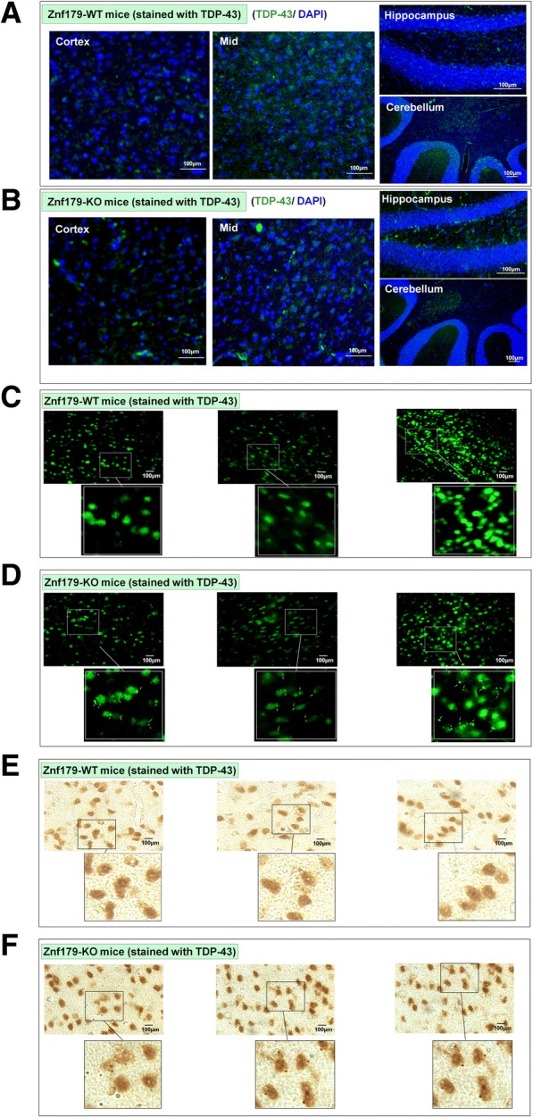
Fig. 7.

Knockout of Znf179 enhances TDP-43 aggregate formation in mice cortex and hippocampus. a and b The brain sections of wild-type (a) or Znf179-knockout mice (b) were stained with anti–TDP-43 antibody (green) and the nuclei were labeled with DAPI (blue). Scale bars = 100 μm. c and d The brain sections of wild-type (c) or Znf179-knockout mice (d) were stained with anti-TDP-43 antibody and the punctate staining of TDP-43 aggregates in the cortex region were indicated by white arrows. Scale bars = 100 μm. e and f The brain sections of wild-type (e) or Znf179-knockout mice (f) were stained with anti-TDP-43 antibody. Scale bars = 100 μm
