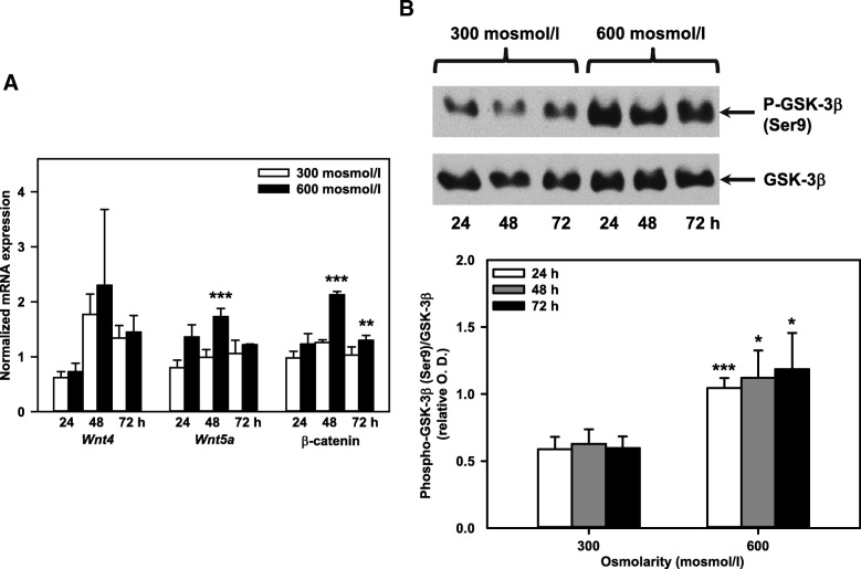
Fig. 4.
Hyperosmolarity increases Wnt/β-catenin signaling in mIMCD3 cells. a Expression levels of Wnt4, Wnt5a and β-catenin mRNA in mIMCD3 cells exposed to norm- or hyperosmotic media for 24-72 h measured by qPCR. The data obtained were normalized to the expression of the reference genes glyceraldehyde-3-phosphate dehydrogenase (Gapdh), β-actin (Actb), and β2-microglobulin (B2m). Means ± SEM of 3-4 experiments are shown. Statistical analysis compares the two osmotic conditions by unpaired t-test. b Expression of GSK-3β and phospho-GSK-3β (Ser9) in mIMCD3 cells was detected by immunoblotting. Optical density (O.D.) of phospho-GSK-3β (Ser9) and GSK-3β signals was analyzed by densitometry using ImageJ software, and relative O.D. was calculated from the ratio of phospho-GSK-3β (Ser9) / GSK-3β. Statistical analysis shows means ± SEM of 4 experiments and compares the two osmotic conditions by unpaired t-test.

