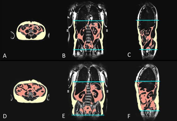
Figure 1.
MRI-based assessment of adipose tissue depots in a 42-year-old male control (a–c); VATvolume 2.8 l, SATvolume 5.8 l, VATarea 89.8 cm2, SATarea 259.4 cm2) and an obese, 57-year-old male with prediabetes (d–f); VATvolume 9.1 l, SATvolume 10.8 l, VATarea 302.3 cm2, SATarea 332.2 cm2). The volumes of the different adipose tissue depots were measured automatically from the diaphragm to the femoral head by employing an in-house algorithm (b–c and e–f). VATarea and SATarea are derived from a single slice on the level of the umbilicus (a, d). (red area = VAT; yellow area = SAT). SAT, subcutaneous adipose tissue; VAT, visceral adipose tissue.

