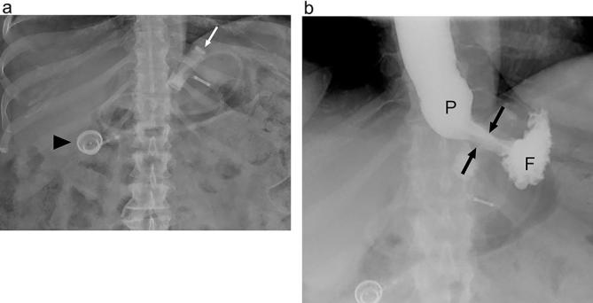
Figure 11.

Expected appearance following LAGB on UGI (a) Supine radiograph shows the expected appearance following LAGB with a band in the left epigastric region (white arrow). Radio-opaque connecting tubing can be assessed as it extends to the injectable port (arrowhead). (b) Supine UGI image acquired while the patient is drinking shows a small pouch (P) with a narrow stoma (arrows) through the band and communicating with the gastric fundus (F). Note that in order to optimally asses the stoma the band must appear linear rather than as a ring shape. LAGB, laparoscopic adjustable gastric banding; UGI, upper gastrointestinal.
