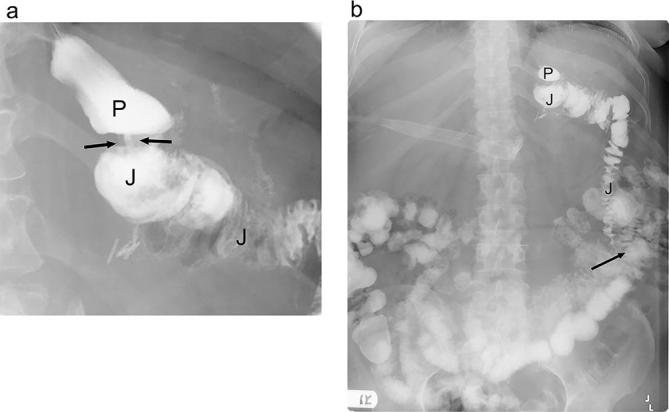
Figure 2.

Expected anatomy following gastric bypass on UGI. (a). Fluoroscopic UGI spot image acquired with the patient in the supine LPO position shows the small gastric pouch (P), narrow gastrojejunal anastomosis (arrows) and adjacent Roux jejunal limb (J). (b). Supine overhead radiograph from UGI shows the gastric pouch (P), Roux jejunal limb (J) and expected location of the left mid-abdominal jejunojejunal anastomosis (arrow). LPO, left posterior oblique; UGI, upper gastrointestinal.
