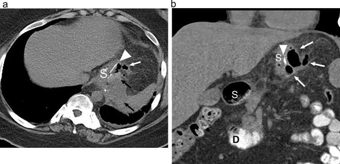
Figure 20.

Leak on CT following SG. (a) Axial and (b) coronal CT images following positive oral contrast administration show an ill-defined fluid collection (black arrows) and extraluminal gas (white arrows) in the left upper quadrant adjacent to the suture line (arrowhead) of the proximal gastric sleeve (S). The fluid collection is of subtle increased density anteriorly due to a small amount of extravasation administered oral contrast. D, duodenum; SG, sleeve gastrectomy.
