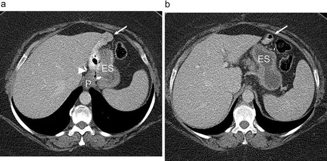
Figure 3.

Expected anatomy following RYGB on CT. (a and b). Axial abdominal CT images acquired with both oral and i.v. contrast show a small gastric pouch (P), gastrojejunal anastomosis (arrowhead), Roux jejunal limb (arrow) and the excluded stomach (ES). Note the opacification of the alimentary jejunal limb (arrow) without opacification of the excluded stomach. RYGB, Roux-en-Y gastric bypass.
