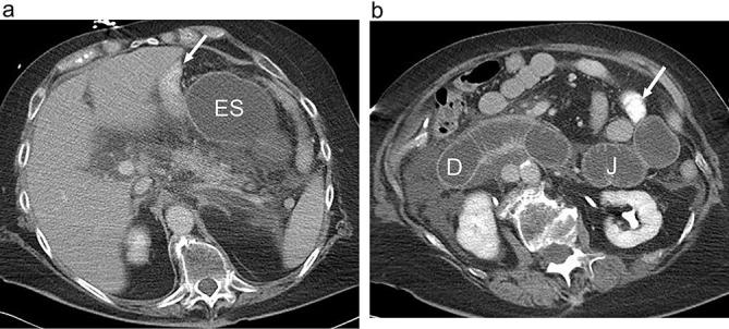
Figure 8.

Small bowel obstruction following RYGB with obstruction of the excluded, biliopancreatic limb. (a and b). Axial CT images following positive oral and i.v. contrast show marked dilatation of the fluid-filled unopacified excluded stomach (ES), duodenum (D) and proximal jejunum (J) (biliopancreatic limb). The opacified alimentary Roux limb is not dilated (arrow). The RYGB anatomy and the jejunal limb must be recognized in order to make the appropriate diagnosis. Distal small bowel is also decompressed. Also note abdominal free fluid. RYGB, Roux-en-Y gastric bypass.
