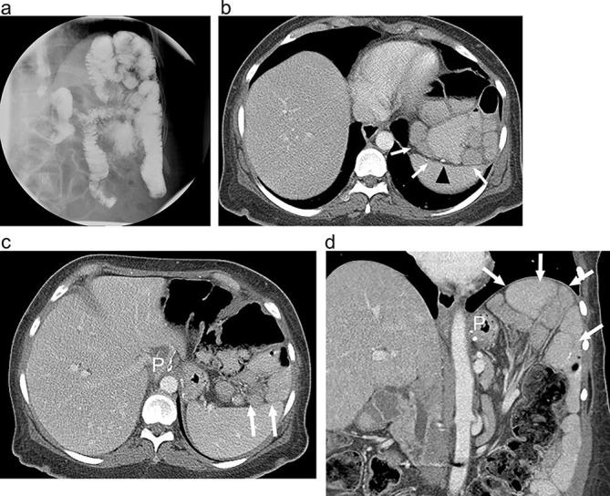
Figure 9.

Internal hernia following RYGB on UGI and CT (a). Supine UGI imaging shows an atypical bowel configuration following RYGB with clustered, displaced small bowel loops (arrows), high in the left upper quadrant, above the gastric pouch (P) and abutting the diaphragm. Small bowel can be seen entering and exiting the clustered segment (arrowhead). (b and c) Axial and (d) coronal CT images with oral and i.v. contrast show RYGB anatomy with clustered displaced small bowel loops high in the left upper quadrant (arrows) above the gastric pouch (P). The jejunojejunal anastomosis is also displaced cephalad (arrowhead) due to internal hernia. Mesenteric vessels are tethered superiorly. At surgery the patient was found to have a large transverse mesocolic internal hernia. RYGB, Roux-en-Y gastric bypass; UGI, upper gastrointestinal.
