Figure 12.
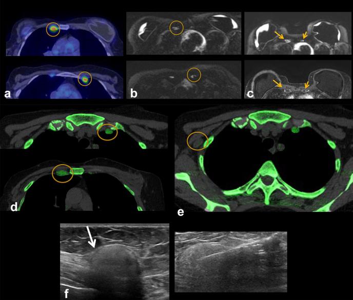
A 50-year-old female with of headaches. Patient had a history of Wilms tumor as a child, status post-nephrectomy and radiation. Patient also had right breast cancer, status post-bilateral mastectomies in over 15 years ago. A PET-CT was obtained to evaluate for recurrent disease. (a) Axial FDG PET-CT: FDG uptake in bilateral IM nodes (circles) and axillary nodes (not shown), highly suspicious for breast cancer metastases. (b) Axial silicone selective MRI shows intact dual lumen silicone implants, with an inner saline shell (low signal intensity) and an outer silicone shell (high signal intensity). High signal is present in IM nodes, suggestive of silicone. (c) Axial silicone selective MRI (top) shows silicone in bilateral IM nodes (arrows). Axial T1 post-contrast MRI (bottom) shows enlarged low signal intensity IM nodes (arrows); silicone appears as low intensity on post-gadolinium images. (d) Axial DECT images show multiple enlarged IM nodes containing silicone as noted by green color mapping (circle) corresponding to PET-CT. (e) Patient also had mild green color mapping in right axillary lymph nodes, suggestive of silicone (circle). (f) Ultrasound of right axillary node demonstrated typical snowstorm appearance of silicone within nodes (arrow). Ultrasound-guided biopsy of the right axillary lymph node was performed for confirmation, which revealed silicone granuloma. Patient underwent explantation of the bilateral breast implants and has done well since, without new pain or symptoms. DECT, dual energy CT; FDG, fludeoxyglucose; IM, .

