Figure 13. .
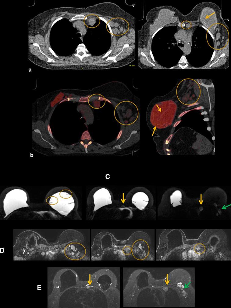
A 58-year-old female presents with tenderness in left chest wall. She has a history of left breast invasive ductal carcinoma, status post-bilateral mastectomy and silicone implant placement 20 years ago, with three implant revisions since. (a) Axial (right) and sagittal (left) non-contrast CT shows enlarged left IM nodes and multiple enlarged left axillary nodes (circles). There are also findings of left implant intracapsular rupture (arrow). (b) Axial (right) and sagittal (left) DECT shows silicone within the left IM and axillary nodes as noted by red color mapping. Intracapsular rupture is also present on DECT as noted by the silicone shell floating within the implant (arrows). DECT, dual energy CT; IM, internal mammary.

