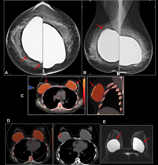
Figure 7. .
A 64-year-old female with bilateral subglandular silicone implant placement over 30 years ago. (a, b) Bilateral screening mammogram shows focal bulges of both implants (arrows), suspicious for implant rupture. C: DECT scanned SUPINE. Some breast tissue not included in scan circle (arrowhead). Sliver of extra-capsular silicone noted outside the calcified capsule bilaterally (arrows). (d) DECT scanned prone. All the tissue included in the scan circle. Radial fold is noted in the right breastarrow), an incidental finding. (e) Silicone sensitive MR sequence shows sliver of silicone outside the fibrous capsule bilaterally (arrows). Findings are consistent with bilateral extracapsular rupture. DECT, dual energy CT.

