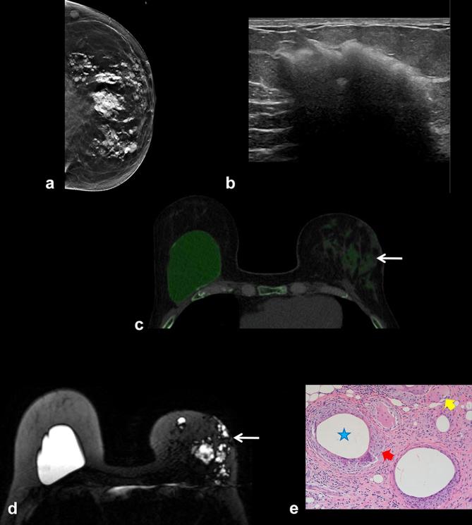
Figure 9.
Patient with extracapsular silicone rupture in the left breast. (a) Mammogram shows high density free silicone throughout the breast parenchyma. (b) Ultrasound shows snowstorm appearance of extracapsular silicone in the breast parenchyma. (c) DECT demonstrates extracapsular silicone in the left breast as green color mapping (arrow). The right breast silicone implant is intact. (d) Silicone sensitive MRI sequence demonstrates ruptured extracapsular silicone as high signal intensity in the left breast (arrow). An intact silicone implant is noted in the contralateral right breast. (e) Pathology demonstrates benign breast parenchyma with dense fibrosis, fat necrosis (star), and foreign body multinucleated giant cell reaction (arrows) consistent with silicone implant rupture. DECT, dual energy CT.

