Figure 11.

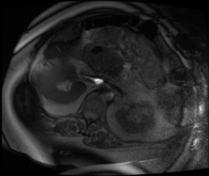
Axial T1 MRI in an obese patient showing wrap around artifact. Images of the anterior pelvis exceeding the field of view are projected along the posterior anatomy. There is also inadequate fat saturation with bands of high signal.

Axial T1 MRI in an obese patient showing wrap around artifact. Images of the anterior pelvis exceeding the field of view are projected along the posterior anatomy. There is also inadequate fat saturation with bands of high signal.