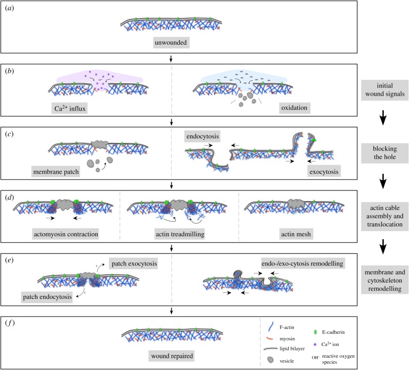
Figure 1.
Schematic of major steps in cell wound repair. (a) Cross section of unwounded cell showing intact plasma membrane and underlying cortical cytoskeleton. (b) Cells with a membrane lesion showing influx of calcium ions (left panel) or reactive oxygen species (right panel) that oxidize cellular components. Influx of these ions each start signalling cascades that initiate cellular wound repair processes. (c) Cells quickly close wounds, either through a vesicular membrane patch (left panel), or by endocytosis/exocytosis of the area of membrane containing the lesion (right panel). (d) Actin based cytoskeleton works to close cellular wounds by an actomyosin contractile ring (left panel), actin treadmilling where the leading edge closest to the wound is the site of F-actin assembly and the trailing edge distal to the wound has F-actin disassembly (middle panel), or by a transient actin patch that accumulates under the membrane lesion (right panel). (e) Cells with repaired wounds must remodel their plasma membrane and cortical cytoskeleton to their pre-wounded state. The fate of the transient membrane patch is not well studied, but it is thought to be removed from the membrane by either endocytosis or exocytosis (left panel). In addition, components of the actin cytoskeleton, which are enriched at the wound site are thought to be either endocytosed or exocytosed in order to return that region to its pre-wounded state (right panel). (f) Cell with completely healed wound—plasma membrane and underlying cortical cytoskeleton have been returned to their unwounded state.

