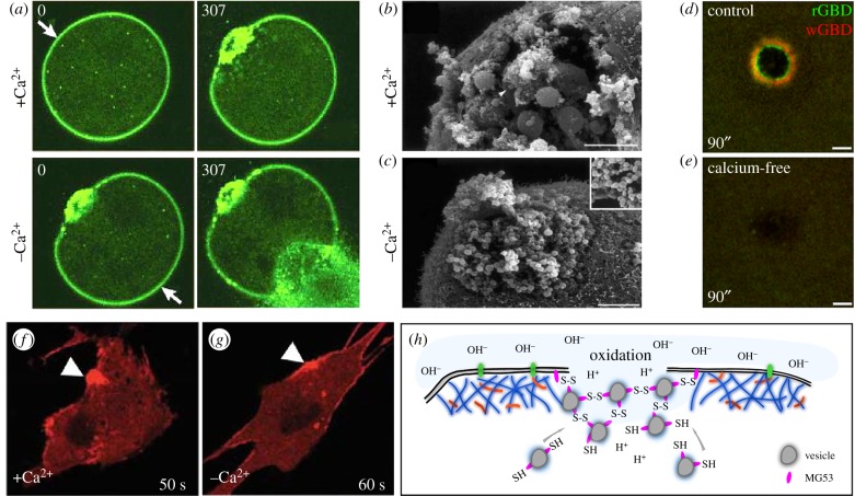
Figure 2.
Calcium influx and oxidation are the most upstream events in cell wound repair. (a) Ca2+ dependence of cell wound repair. An FM1-43 labelled sea urchin egg was wounded twice with a laser. The first wound (arrow; upper left panel) was generated in the presence of calcium and exhibits a robust repair response (upper right panel). The sea urchin egg was moved to calcium-free sea water and a second wound (arrow; lower left panel) was then generated. In the absence of calcium no repair is initiated and cytoplasm can be seen flowing out of the wound (lower right panel). Adapted by permission from Springer Nature and Copyright Clearance Center: McNeil & Kirchhausen [4] (Copyright © 2005). (b,c) Scanning electron micrographs of sea urchin eggs in the presence (b) or absence (c) of calcium upon wounding. Wounds were generated mechanically using a needle. In the presence of calcium, vesicles (arrowhead) are recruited to the wound where they fuse to each other resulting in the formation of large vesicles. In the absence of calcium, no large vesicles are formed. Adapted by permission from Springer Nature and Copyright Clearance Center: McNeil & Baker [47] (Copyright © 2001). (d,e) Recruitment of activated Rho family GTPases in Xenopus oocytes upon wounding in the presence (d) or absence (e) of calcium. Activity biosensors for Rho (rGBD) and Cdc42 (wGBD) only exhibit distinct concentric ring patterns around the wound in the presence of calcium. Republished with permission of The Rockefeller University Press, from Benink & Bement; permission conveyed through Copyright Clearance Center, Inc. [24] (Copyright © 2005). (f,g) MG53 protein tethered to vesicles and membrane accumulates at wounds (arrowheads) even in the absence of calcium. Adapted by permission from Springer Nature and Copyright Clearance Center: Cai et al. [13] (Copyright © 2009). (h) Schematic depicting the role of oxidation in cell wound repair. Vesicles coated with MG53 protein are recruited to wounds where the MG53 proteins attach to each other through oxidation-dependent disulfide bond formation.

